Glossary

What is Electromyography?

Electromyography is a diagnostic test that measures the electrical activity of muscles and the nerves controlling them. Using small electrodes, it records how muscles respond to nerve signals, helping identify nerve or muscle disorders, injuries. Or conditions like carpal tunnel syndrome or sciatica.

Reviewed by ChiropractorMcDonough.comSources reviewed: American Association of Neuromuscular & Electrodiagnostic Medicine, National Institute of Neurological Disorders and Stroke

Quick Facts About Electromyography

Category

Diagnostic test

Used for

Identifying nerve or muscle disorders

Common confusion

Often mistaken for nerve conduction studies, which measure nerve speed separately

Also called

EMG, Muscle electrical activity test

Often discussed with

Chiropractic Consultation and Diagnosis, Whiplash Injury Treatment

Key Takeaways About Electromyography

Understanding Electromyography

Electromyography in Chiropractor: Electromyography is a diagnostic test that measures the electrical activity of muscles—v...

Electromyography, often called EMG, is a medical test that checks the health of muscles and the nerves that control them. When muscles move, they produce tiny electrical signals. An EMG machine records these signals to see if muscles are responding correctly to nerve messages. If the signals are weak or missing, it could mean there is a problem with the nerves or muscles.

Related glossary terms: Nerve Compression, Chiropractic Neurology, Orthopedic Tests.

This test is commonly used to diagnose conditions that cause pain, weakness. Or numbness. For example, if someone has sciatica—a condition where a pinched nerve causes leg pain—an EMG can help pinpoint which nerve is affected. It can also detect muscle diseases, nerve injuries. Or disorders like carpal tunnel syndrome. The test is usually done in a clinic or hospital by a trained specialist.

How Electromyography Works?

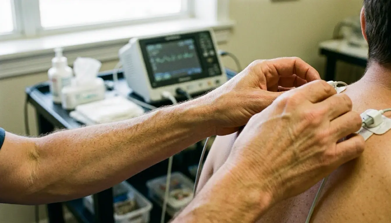
During an EMG, small electrodes are used to pick up electrical activity. Surface electrodes are placed on the skin over a muscle. While needle electrodes are inserted directly into the muscle. The patient may be asked to relax or gently contract the muscle while the machine records the signals. These signals appear as waves on a screen or printout, showing how strong and frequent the electrical activity is.

The test has two main parts. First, the electrodes record activity while the muscle is at rest. Healthy muscles should show little to no electrical activity when relaxed. The result matters. Second, the patient is asked to contract the muscle, like bending an arm. The machine records how the signals change during movement. If the signals are abnormal, it can help the doctor determine if the problem is in the muscle, the nerve. Or the connection between them.

Why Electromyography Matters?

How Electromyography applies to Chiropractor services in McDonough, United States—practical illustration

Electromyography is important because it provides detailed information that other tests can't. For example, an X-ray can show bone problems. But it cannot detect nerve or muscle issues. An EMG helps doctors confirm a diagnosis, plan treatment. Or monitor how well a condition is improving. Without this test, some nerve or muscle disorders might go undiagnosed or be treated incorrectly.

The results of an EMG can guide decisions about physical therapy, medication. Or even surgery. For instance, if the test shows severe nerve damage, a doctor might recommend surgery to relieve pressure on the nerve. If the problem is mild, physical therapy or lifestyle changes might be enough. This makes EMG a valuable tool for both patients and healthcare providers.

When Electromyography Matters Most?

Electromyography is most useful when symptoms suggest a nerve or muscle problem. Common signs include muscle weakness, tingling, numbness. Or pain that doesn't go away. It's often ordered for people with conditions like sciatica, carpal tunnel syndrome. Or muscle diseases such as muscular dystrophy. It can also help diagnose nerve damage caused by injuries, diabetes. Or other chronic illnesses.

The test is also helpful for tracking progress during treatment. For example, if someone is recovering from a nerve injury, repeated EMGs can show whether the nerve is healing. This helps doctors adjust treatment plans as needed. In some cases, an EMG is used to rule out certain conditions, giving patients clear next steps when other tests come back normal.

How to Evaluate Electromyography?

Related Concepts Compared

Electromyography vs. Nerve Conduction Study

A nerve conduction study measures how fast electrical signals travel through nerves. While electromyography focuses on muscle response to those signals.

Electromyography vs. MRI

An MRI shows detailed images of soft tissues and bones but cannot measure electrical activity like an EMG.

Electromyography vs. Ultrasound

Ultrasound creates images of muscles and nerves but does not record electrical signals like an EMG.

Expert Note

Electromyography is highly sensitive to nerve and muscle function but requires careful interpretation. Abnormal findings should always be correlated with clinical symptoms and other diagnostic tests to avoid misdiagnosis.

Common Mistakes or Myths About Electromyography

  • Assuming an EMG is painful—while needle electrodes may cause slight discomfort, most patients tolerate the test well.
  • Confusing EMG with nerve conduction studies—both are often done together but measure different things.
  • Thinking EMG is only for severe conditions—it can also diagnose mild nerve or muscle problems early.
  • Ignoring preparation instructions, like avoiding lotions or oils on the skin before the test.

Electromyography in Practice: A Real-World Example

A patient visits a chiropractor with persistent leg pain and numbness. After a physical exam, the chiropractor refers the patient for an EMG. The test reveals a pinched nerve in the lower back, confirming sciatica. This helps the chiropractor create a targeted treatment plan, including spinal adjustments and physical therapy.

Related Services

Related Terms

Nerve Compression

Nerve Compression is a condition where a nerve is pressed or squeezed by surrounding tissues, such as bones, muscles, tendons. Or cartilage. This pressure disrupts the nerve’s normal function, often causing pain, numbness, tingling. Or weakness in the affected area. Common sites include the spine, wrists. And elbows. And symptoms may worsen with movement or prolonged pressure.

Chiropractic Neurology

Chiropractic Neurology is a specialized branch of chiropractic care that focuses on the relationship between the nervous system and spinal function. It uses non-invasive techniques to assess and improve neurological health, helping patients with conditions like balance disorders, chronic pain. And brain-body coordination issues by evaluating nerve pathways and brain function.

Orthopedic Tests

Orthopedic Tests are hands-on examinations chiropractors and doctors use to diagnose muscle, joint, nerve. Or bone problems. These tests involve specific movements, pressure. Or positions that reproduce symptoms, helping identify the exact source of pain or dysfunction without imaging. They're quick, non-invasive.

Manual Therapy

Manual Therapy is a hands-on clinical approach used by chiropractors, physical therapists. And other healthcare providers to diagnose and treat musculoskeletal conditions. Manual Therapy includes techniques like joint mobilization, soft tissue manipulation, stretching. And spinal adjustments to reduce pain, improve mobility. And restore function without surgery or medication.

ChiropractorMcDonough.com

Have Questions About Electromyography?

Contact ChiropractorMcDonough.com for practical guidance on Electromyography and related chiropractor work in McDonough.

+1 770-637-6261